Understanding the periodontal ligament, a vital component of dental anatomy, is intrinsically linked to the health and integrity of the lamina dura function and structure. The alveolar bone, specifically its inner socket lining, is where we find the lamina dura, observable through tools such as a dental radiograph. Oral health professionals frequently assess the appearance of the lamina dura because changes in its density and thickness often indicate underlying dental or systemic conditions, making a thorough understanding of lamina dura function and structure essential for accurate diagnosis.
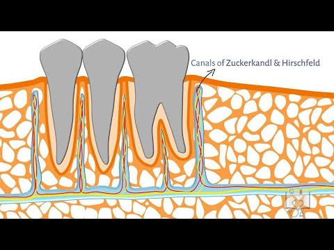
Image taken from the YouTube channel Doctoropsy , from the video titled Alveolar Bone- Structure .
Understanding the Lamina Dura: Its Role and Anatomy
The lamina dura is a critical, though often overlooked, component of dental health. This article provides a straightforward explanation of the lamina dura function and structure, clarifying its significance in maintaining teeth and overall oral well-being.
What is the Lamina Dura?
The lamina dura is a thin, compact layer of bone that lines the tooth socket (alveolus) in the jawbone. It’s visible on dental X-rays as a radiopaque (lighter) line surrounding the tooth root. This line represents the bone that directly supports the tooth. It’s vital for proper tooth anchorage and response to changes in bite force.
Composition of the Lamina Dura
The lamina dura is primarily composed of dense, cortical bone. This type of bone is strong and relatively dense, providing significant support. Key components include:
- Calcium Phosphate: The primary mineral constituent, contributing to the bone’s hardness.
- Collagen Fibers: These provide a flexible framework within the bone matrix.
- Osteocytes: Bone cells embedded within the lamina dura, responsible for maintaining bone tissue.
- Blood Vessels: Provide nourishment and facilitate bone remodeling.
- Nerve Fibers: Contribute to sensory feedback and bone maintenance.
The Function of the Lamina Dura
The lamina dura performs several essential functions crucial for tooth health and stability.
Tooth Anchorage and Support
The primary role of the lamina dura is to provide a rigid attachment point for the periodontal ligament (PDL). The PDL connects the tooth root to the alveolar bone, and the lamina dura acts as the bony anchor for these fibers. This anchorage is essential for:
- Resisting forces during chewing: The lamina dura helps distribute occlusal forces, preventing excessive stress on the tooth.
- Maintaining tooth position: It helps keep the tooth firmly in its socket, preventing unwanted movement.
- Protecting the tooth root: It acts as a protective barrier around the tooth root.
Response to Occlusal Forces and Bone Remodeling
The lamina dura is not static. It actively remodels itself in response to changes in bite force and other stimuli. This remodeling process is facilitated by specialized cells.
- Osteoblasts: Cells that build new bone tissue, thickening the lamina dura in areas subjected to increased stress.
- Osteoclasts: Cells that resorb (break down) bone tissue, thinning the lamina dura in areas of reduced stress or inflammation.
This dynamic process allows the lamina dura to adapt to changing conditions, ensuring optimal tooth support.
Indication of Oral Health
The appearance of the lamina dura on dental X-rays is a valuable indicator of oral health. Changes in its thickness or density can signal underlying dental problems.
Interpreting the Lamina Dura on X-Rays
Dental professionals carefully examine the lamina dura on X-rays to assess its health.
Normal Appearance
A healthy lamina dura appears as a continuous, radiopaque (white) line around the tooth root. Its thickness should be relatively uniform.
Abnormal Appearance
Several abnormalities can indicate dental problems. Some of them are:
- Loss of the Lamina Dura: Disappearance of the lamina dura, often indicating advanced periodontal disease or periapical pathology (infection at the tip of the root).
- Thickening of the Lamina Dura: Thickening can suggest increased occlusal forces or bruxism (teeth grinding).
- Localized Thinning: May indicate localized inflammation or early periodontal disease.
- Irregular Density: Uneven density could signify metabolic bone disease or other systemic conditions.
| Appearance | Possible Cause |
|---|---|
| Loss | Periodontal Disease, Periapical Lesion |
| Thickening | Bruxism, Increased Occlusal Forces |
| Localized Thinning | Localized Inflammation, Early Periodontal Disease |
| Irregular Density | Metabolic Bone Disease, Systemic Conditions |
It’s important to note that X-ray interpretation requires professional expertise.
Factors Affecting the Lamina Dura
Various factors can influence the health and appearance of the lamina dura.
- Periodontal Disease: Inflammation and bone loss associated with periodontal disease can directly affect the lamina dura.
- Occlusal Trauma: Excessive biting forces can cause thickening or damage to the lamina dura.
- Systemic Diseases: Conditions like osteoporosis or hyperparathyroidism can alter bone metabolism, affecting the lamina dura.
- Medications: Certain medications, such as bisphosphonates, can impact bone remodeling and influence the lamina dura.
FAQs: Lamina Dura – Function & Structure
Here are some frequently asked questions to help you understand the lamina dura better.
What exactly is the lamina dura?
The lamina dura is a thin, dense layer of bone that lines the tooth socket (alveolus). It’s part of the alveolar bone and is visible on dental X-rays. Understanding the lamina dura function and structure is key to identifying healthy teeth and bone.
What is the main job of the lamina dura?
The primary function of the lamina dura is to provide a hard, bony surface for the attachment of periodontal ligament fibers. These fibers connect the tooth root to the alveolar bone, securing the tooth in place. Therefore, proper lamina dura function and structure is crucial for tooth stability.
How does the lamina dura appear on an X-ray?
On a dental X-ray, the lamina dura appears as a distinct white line surrounding the tooth root. Its density reflects its mineral content. Changes in its appearance can indicate underlying dental or bone issues, emphasizing the importance of lamina dura function and structure assessment.
What does it mean if the lamina dura doesn’t look normal on an X-ray?
An abnormal-looking lamina dura on an X-ray can indicate a variety of problems, such as infection, trauma, or even systemic diseases. A dentist can interpret these changes, because lamina dura function and structure appearance on the X-ray gives important diagnostic information. It is important that the dentist assess the context with all other examination findings.
So, there you have it! Hopefully, this explanation of lamina dura function and structure cleared things up. Keep that smile healthy!